Nail Anatomy
Nail Anatomy
1. Overview
The human fingernail is a hard, keratinous structure at the tip of each finger. [1] It protects the fingertip, helps with tactile sensation and precision tasks, and plays an important aesthetic role. For manicure and nail extension work, several parts of the nail unit are especially relevant.
2. Anatomy Parts
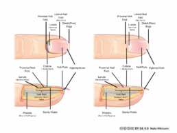
The nail unit (also called the nail apparatus) consists of several interconnected parts. Each has a unique structure and purpose that contributes to the nail’s appearance, strength, and growth.

2.1 Nail plate (corpus unguis)
The nail plate is the hard, visible part of the nail. It is made of compact layers of keratinized cells, a tough protein similar to what forms hair. [2] These cells are produced by the nail matrix and slide forward as new layers grow beneath them. [1]
Although it looks solid, the nail plate is slightly porous and flexible due to it consisting it of keratin [3], allowing it to bend without breaking.
2.2 Nail Bed (Leticulum unguis)
The nail bed lies directly beneath the nail plate and contains numerous small blood vessels, giving the nail its pinkish color. It provides structural support and helps the nail plate adhere to the finger. [4]
Because of its rich vascular network, injuries to the nail bed often cause pain and bruising, and can lead to temporary nail changes.
2.3 Nail Matrix (Matrix unguis)
The matrix is the living growth center of the nail, located beneath the skin at the base of the nail plate. Here, specialized cells divide and harden into keratin, forming the new nail. [5]
Damage to the matrix, e.g. through trauma, infection, or improper cosmetic procedures, can cause permanent nail deformities, such as ridges or uneven growth.
2.4 Lunula
The lunula (Latin for “little moon”) is the whitish, crescent-shaped area visible at the base of some nails. It represents the visible portion of the matrix. [4]
Its size and visibility vary from person to person and are not necessarily indicators of health.
2.5 Cuticle (Eponychium) and Nail Folds (Paronychium)
The cuticle, also known as the eponychium, is a thin layer of skin that overlaps the base of the nail plate. It forms a protective seal between the nail plate and the surrounding skin, preventing bacteria and fungi from entering the nail matrix and protecting it from UV light. [4]
The lateral nail folds (paronychium) border the sides of the nail and serve a similar protective function. [2] Cutting or aggressively removing the cuticle increases the risk of infection and inflammation. [4]
3. Nail Properties
3.1 Nail Function
The nail is not just decorative but it plays several important protective and sensory roles:
-
Protection: Shields the sensitive fingertip and underlying tissue from injury. [4]
-
Tactile enhancement: Works together with the fingertip’s nerves to improve fine touch and precision. [4]
-
Support and grip: Provides counterpressure for grasping, scratching, and manipulating small objects. [4]
-
Aesthetic expression: Serves as a visible reflection of personal care, culture, and identity.
3.2 Nail Growth
Nails grow continuously throughout life. On average, a fingernail grows about 0.1 mm per day, or roughly 3 mm per month. [1]
Complete regrowth of a fingernail takes four to six months [4], depending on individual factors such as:
-
Age: Younger people tend to have faster nail growth.
-
Season: Growth is often faster in summer than in winter.
-
Health and Nutrition: Illness, poor circulation, or nutrient deficiencies (especially biotin and iron) can slow nail production.
-
Dominant Hand: Nails on the dominant hand often grow faster due to increased blood flow and use.
Fun fact: Toenails grow much more slowly, about one-third the speed of fingernails. [1]
3.3 Nail Permeability (Porosity)
Although the nail plate appears hard and impermeable, it actually allows limited passage of water and certain substances.
The keratin that forms the nail plate is more permeable to moisture than the surrounding skin, meaning water and small molecules can pass through it more easily.
On average, the nail substance itself contains about 7–12% water under normal conditions. [6]
When exposed to moisture for prolonged periods, the nail plate absorbs water and swells slightly. This temporary swelling softens the keratin structure and changes the nail’s flexibility and surface texture. [6]
Because of this property, chemicals and even medications can also penetrate the nail plate to a limited extent, a fact that is important for both cosmetic applications (like nail treatments or polishes) and medical therapies (such as antifungal agents).
4. Common Nail Changes and What They Mean
There are many different diseases and disorders that can cause changes in the nails.
| Change | Description | Possible Cause |
|---|---|---|
| Longitudinal ridges | Fine vertical lines along the nail | Normal with age, dryness |
| Transverse ridges (Beau’s lines) | Horizontal grooves | Temporary growth interruption (illness, stress) |
| Brittle nails | Splitting or breaking easily | Excessive moisture, harsh chemicals, dehydration |
| White spots (Leukonychia) | Small white dots on the plate | Minor trauma to the matrix |
| Yellow discoloration | Yellow or brownish tint | Smoking, nail polish pigments, fungal infection |
| Pitting | Small dents or holes | Psoriasis, eczema, trauma |
Note: Persistent or painful nail changes should always be evaluated by a healthcare professional or dermatologist.
Sources & Links
[1] Altmeyers Encyclopedia – Nail – Prof. Dr. med. Peter Altmeyer – https://www.altmeyers.org/en/dermatology/nail-120404
[2] National Library of Medicine of the US – nail anatomy –
https://www.ncbi.nlm.nih.gov/books/NBK513133//
[3] UC San Diego – Keratin: Structure, mechanical properties, occurrence in biological organisms, and efforts at bioinspiration – Wang, Bin; Yang, Wen; McKittrick, Joanna; et al. – https://escholarship.org/content/qt5sb7q6jp/qt5sb7q6jp.pdf?t=o9xpu0
[4] Cleveland Clinic – Nails (Fingernails, Toenails) –
https://my.clevelandclinic.org/health/body/nail-anatomy
[5] Biology Online – Nail Matrix –
https://www.biologyonline.com/dictionary/nail-matrix
[6] Doccheck Flexikon – Nagel –
https://flexikon.doccheck.com/de/Nagel